



Since scientists discovered jumping genes in maize in 1950, they have known that bits of DNA can move around the genome, mostly within the cell nucleus. Scientists have also found evidence of genes having jumped across species.
Now, researchers at the Max Planck Institute for Marine Microbiology in Germany have reported a remarkable observation: they have visually tracked an RNA molecule, called an intron, as it jumped from one organism into another.
This is remarkable because the scientists literally caught the gene as it jumped across species, without the help of any courier.

Jumping genes — technically now called transposable elements — are responsible for the spread of antibiotic resistance and in the development of several cancers. Scientists are also studying circular RNA molecules, like the one derived from the jumping intron, as next generation vaccine platforms. Understanding the lives of jumping genes can thus have profound implications for evolution and ultimately to medicine and biotechnology.
A tiny predator
Microbial communities that occur naturally are very difficult to nurture in large numbers in a lab because they will be isolated from their surroundings. So the team at Jens Harder’s laboratory seeded a lab culture with a small amount of wastewater from a local sewage treatment plant.
Over time, this culture of microbes came to consist of several members of archaea and bacteria. Like bacteria, archaea are another domain of life, and are also microscopic.
Microbes of both these domains lack a true nucleus. Instead, their nucleus lies in the cytoplasm itself.
The team focused on two members of this community. One member is the methane-producing archaeon, Methanothrix soehngenii. The other is a bacterium so small that it is called an ultramicrobacterium. The team named it Velamenicoccus archaeovorus. It is a predator: it attaches to the surface of the archaeon, breaks apart its cell membrane, and kills it.

The rough draft
It is impractical to investigate each member of a highly diverse microbial community separately, so scientists study them en masse. In the study, the researchers identified the RNA molecules in the members’ ribosomes. The ribosome is each cell’s protein-making factory.
The ribosomes in all cells of all life forms have RNAs called ribosomal RNAs (rRNA). All life forms have rRNA that perform the same function. However, each species has a unique sequence of bases that can be used to identify the species. This way, the scientists determined the composition of their lab culture.
In the ultramicrobacterium, one type of rRNA called 23S is initially made as a precursor — like a rough draft, something the cell has to edit further. This editing takes the form of losing a particular sequence of bases called the intron. Essentially, the intron is cut out of the precursor.
The intron itself is a special kind of RNA molecule with two unique roles: it cuts itself out of the precursor rRNA and joins its own ends up to acquire a circular structure. And it contains information that can allow it to ‘jump’.
The team also analysed the rRNA sequences of the ultramicrobacterium from a previous study. This revealed that the introns were highly efficient at their tasks: they rarely failed.

Tracking by eye
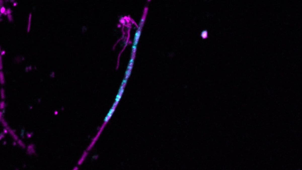
Finally, the team decided to check visually whether the intron of the 23S rRNA of the predator jumped to the prey as the two interacted.
Other scientists at the same Max Planck Institute had already found, and perfected, a way to track RNA visually. It consisted of tracking the RNA using a probe with a pair of special features. One, it was designed to home in on the intron (based on its sequence) and latch on to it. Two, the probe was attached to a fluorescent label — a chemical compound that glows when seen through a fluorescence microscope.
The team modified the probe so that its label would glow green in live cells but yellow in dead cells — either way only if the intron was present.
The team also could tell which cells the intron was by using the difference in appearance of the microbes. V. archaeovorus appears as small spots while Methanotrix soehngenii appears as filaments.
This way, the team detected the intron in Methanotrix soehngenii cells. Specifically, there was a faint yellow glow in the filamentous structures. This meant the cells in which the intron was present were dead.
Clearly, the intron RNA had exited the predator cells and entered the prey cells.

Truly remarkable
RNA molecules are famously unstable, so how did the introns survive this jump? The researchers proposed that the intron’s self-circularisation could be the reason. The circular shape of the RNA molecule allows it to resist being degraded by a cell’s enzymes. The ultramicrobacterium’s genome also encodes an enzyme that could have helped the intron insert itself into foreign DNA.
There are still some unanswered questions. For example, does evolution intend for introns to be integrated into prey DNA? In the experiment, the intron actually jumped into dead cells. Why? Or, alternatively, did the intron kill the cells?
Visual confirmation that an intron can jump across two different domains of life is truly remarkable. Still, let us remember that it happened in a laboratory culture (even if there is indirect evidence that introns do jump in the real world as well).
Living beings sometimes swap genes with viruses, which are the essential couriers of gene transfer. This is known as horizontal gene transfer. The new study, however, has reported the first direct visual evidence of horizontal gene transfer across species without a courier.
Studying this phenomenon helps scientists understand how organisms rapidly acquire new traits, such as antibiotic resistance or new metabolic abilities. It also reveals how genes move across species, potentially reshaping ecosystems and driving the rise of new disease-causing microbes.
S. Swaminathan is a retired professor of biology (BITS-Pilani, Hyderabad) and a former scientist (ICGEB, New Delhi).
Published – July 15, 2026 09:00 am IST
